

Our Projects
01
Automated radiation-free assessment of scoliosis using artificial intelligence and 3D ultrasound imaging
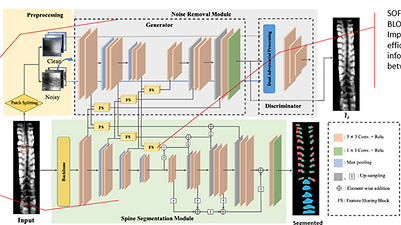
Adolescent idiopathic scoliosis (AIS) affects between 2-4% of adolescents in the general population. Curve progression is the most probable occurrence among teenagers with AIS, the regular observation is essential for monitoring curve progression. If not appropriately treated, progressive scoliosis may cause constant back pain, poor posture, shoulder humping, breathing problems, or even physical disability for life. Sometimes may require surgery. Thus, the early detection of scoliosis is critical to providing effective clinical treatments to prevent scoliosis progression during growth. Therefore, large-scale school scoliosis screening is part of the national health program in many countries. A common test used to screen for scoliosis is called the “Adams forward bending test”. Such screening methods often identify many false positives, thereby causing unnecessary anxiety amongst subjects and their parents, in addition to burdening the healthcare system. The gold standard for identifying and monitoring AIS has been standing anteroposterior and lateral X-ray imaging. A teenager with scoliosis may receive more than 20 X-rays over the course of their treatment. It will significantly pose health risks by repeated exposure to radiation. Also, people in rural areas can have limited access to X-ray imaging facilities.
In this project, an automatic ultrasound-based radiation-free 3D screening tool will be developed for large-scale diagnosis of AIS. Advanced artificial intelligence (AI) techniques will be developed to tackle the challenges associated with accurately analysing low-quality ultrasound images. An innovative and effective 2.5D sequencing approach will be developed to capture the 3D inter-slice information to boost accuracy without adding extra computational burden. The resultant low-cost automatic scoliosis assessment system can be used for large-scale school scoliosis screening across Australia, with AI breakthrough revitalising ultrasound based health care.

02
Deep Classification of Electroencephalography Signals for Brain-Computer Interfacing Applications
The proposal outlines a research initiative focused on enhancing the classification of electroencephalography (EEG) signals using computational intelligence (CI) techniques for brain-computer interface (BCI) applications. The aims encompass employing CI technologies to improve EEG signal classification accuracy, developing variant-topologies for fusion-based convolutional neural network (CNN) classifiers, and introducing automated meta-heuristic designs for these networks. Stability analysis of the created networks will be performed to optimize the number of reliable classes, crucial for enhancing BCI reliability. Additionally, the project aims to develop innovative applications for the efficient utilization of the model in real-world scenarios, potentially advancing fields such as medicine, robotics, and military technology. By addressing these objectives, the research endeavors to enhance the performance, reliability, and practical applicability of EEG-based BCIs, paving the way for their widespread adoption and utilization across various domains.


03
Data-Centric AI for Large-Scale Clinical Annotation Recovery in Echocardiography
The rapid advancement of foundation models in medical imaging has highlighted that data quality and annotation availability are often more important than model architecture itself. In echocardiography, millions of clinical studies are generated annually, containing valuable anatomical measurements and expert annotations created by sonographers during routine patient examinations. However, much of this information remains inaccessible for artificial intelligence development because measurement calipers, landmarks, and clinical labels are embedded directly within image pixels rather than stored as structured annotations. As a result, a vast amount of clinically validated knowledge remains locked within existing archives and cannot be readily used to train modern AI systems.
The aim of this PhD project is to develop a comprehensive Data-Centric AI framework that automatically discovers, recovers, validates, and curates clinically meaningful annotations from large-scale echocardiography archives to create foundation-scale datasets for next-generation cardiac imaging AI systems. Rather than focusing on developing new neural network architectures, the research emphasises the systematic improvement of data quality, annotation reliability, and dataset scalability, recognising that high-quality data is the key enabler for future foundation models and vision-language models in healthcare

04
Generative AI for Ultrasound – Reconstruction, Restoration, and Synthetic Data Generation
Ultrasound imaging is one of the most widely used diagnostic modalities in modern healthcare, yet the development of artificial intelligence systems for ultrasound remains constrained by limited annotated data, image artefacts, vendor variability, and the presence of clinical overlays such as measurement calipers and textual annotations. Unlike natural images, ultrasound images contain unique speckle patterns and acoustic characteristics that are essential for clinical interpretation. Existing generative AI approaches developed for natural images, CT, or MRI often fail to preserve these ultrasound-specific properties, limiting their applicability in clinical practice. This PhD project aims to establish a new research direction in Generative AI for Ultrasound by developing advanced generative models capable of reconstructing obscured anatomical structures, restoring clinically realistic ultrasound images, and generating high-quality synthetic datasets for foundation model development.
Aim & Objectives: The main aim of this project is to develop ultrasound-specific generative artificial intelligence frameworks that can reconstruct, restore, and synthesize clinically realistic ultrasound images while preserving anatomical integrity and ultrasound physics characteristics. The research will focus on creating a complete pipeline that transforms annotated clinical images into clean, AI-ready datasets and generates additional synthetic data to support the training of next-generation medical foundation models.

05
Foundation Models for Echocardiography – Towards General-Purpose AI for Cardiac Ultrasound
Echocardiography is one of the most important imaging modalities for assessing cardiac anatomy and function, yet most existing artificial intelligence solutions remain highly task-specific, focusing on individual applications such as chamber segmentation, landmark detection, ejection fraction estimation, or disease classification. These models typically require large amounts of labelled data for each task and often struggle to generalise across different imaging views, vendors, patient populations, and clinical scenarios. Recent advances in foundation models have demonstrated the potential of large-scale pretraining to create general-purpose AI systems capable of adapting to multiple downstream tasks with minimal additional supervision. However, the development of foundation models for echocardiography remains in its infancy due to the scarcity of large-scale, high-quality annotated datasets and the unique challenges associated with ultrasound imaging.
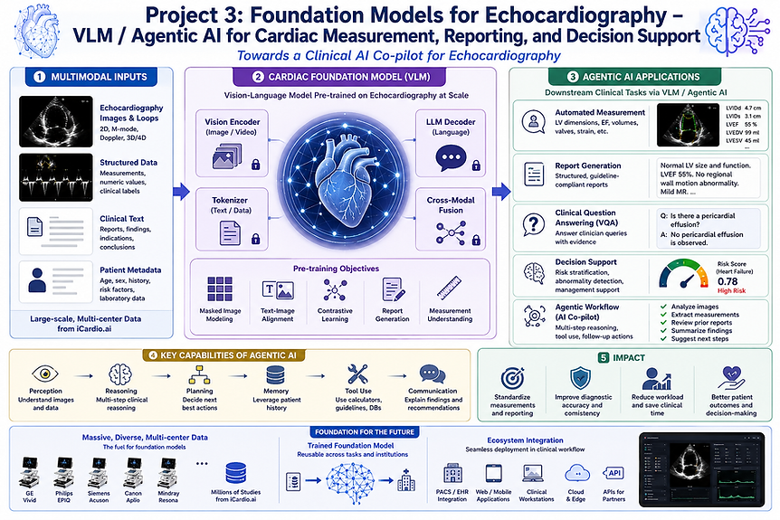
The aim of this PhD project is to develop a foundation model for echocardiography that learns comprehensive anatomical, structural, and functional representations from large-scale multimodal ultrasound data, enabling generalisable cardiac image understanding, automated measurement, and clinical decision support across a wide range of echocardiographic tasks. The research seeks to move beyond conventional task-specific models by establishing a unified framework that can perform multiple cardiac imaging tasks using a single pretrained model.
06
TrustMed-VLM: A Trustworthy Vision–Language and Large Language Model Framework for Clinically Reliable Medical Imaging Analysis
TrustMed-VLM is a novel trustworthy Vision–Language and Large Language Model framework designed to address the key limitations that currently hinder the clinical adoption of multimodal AI systems in medical imaging. Existing VLM–LLM models often suffer from hallucination, weak visual localisation, limited generalisation to medical imaging domains, and an inability to effectively process multidimensional imaging data such as videos and volumetric scans. To overcome these challenges, TrustMed-VLM integrates domain-adaptive medical representation learning, forced visual grounding, retrieval-augmented clinical reasoning, and trust-aware reliability assessment within a unified architecture.
The framework supports multiple imaging modalities, including X-ray, ultrasound, echocardiography, CT, and MRI, while extending beyond conventional two-dimensional analysis to incorporate temporal and volumetric reasoning. A key innovation is the use of forced visual grounding, which requires every generated clinical conclusion to be explicitly linked to corresponding anatomical structures, lesions, or imaging evidence, thereby improving explainability and reducing hallucinations. Retrieval-augmented generation further enhances factual consistency by incorporating external clinical knowledge, guidelines, and historical cases during reasoning. In addition, a dedicated trust assessment module evaluates localisation confidence, uncertainty, and reasoning consistency before producing outputs. By combining trustworthy reasoning, visual evidence verification, and multidimensional image understanding, TrustMed-VLM aims to provide clinically reliable decision support, report generation, and diagnostic assistance for real-world healthcare applications.